ABtest-IA

ABtest-IA is a colorimetric sandwich ELISA that requires 300 microliters of plasma for the determination of Aβ40 and Aβ42.

Analytical Validation

The assay is fully validated from an analytical point of view according to EMA and FDA recommendations and is CE marked in the European Union and Investigational Research Only (IUO) in the rest of the world.

Clinical Results

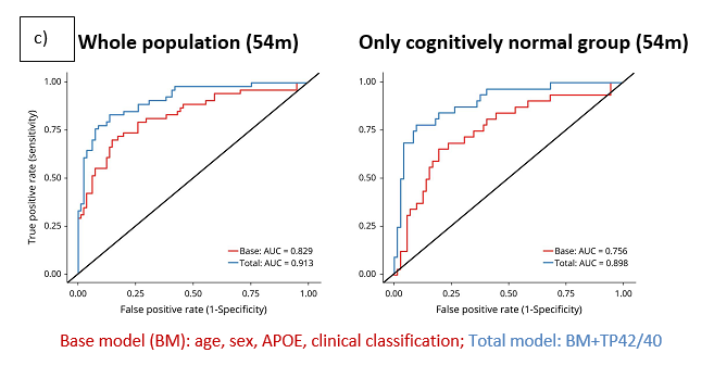
The plasma Aβ42/Aβ40 ratio has been predictive of Amyloid-PET status with areas under the ROC curve (AUCROC) ranging from 0.77 to 0.88 in different cohorts (0.78 to 0.96 when the APOE genotype is included in the predictor model).

Recruitment Hypothetical Scenario

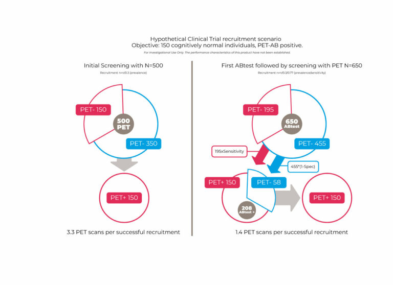
In a hypothetical recruitment scenario for a clinical trial of cognitively healthy individuals with confirmed amyloid brain pathology, pre-screening with ABtest would save up to 60% of the required amyloid PET scans (or CSF analysis).
Because of its capacity to analyse large numbers of samples in a short time and at low cost, ABtest-IA is particularly suitable for the study of previously stored specimen collections.

ANALYTICAL VALIDATION

ABtest40 and ABtest42 accuracy for plasma samples of 5 different concentrations. Each point represents the relative error (RE, %) of each of the measurements made. The dotted lines indicate the limit of compliance established by the validation guidelines (± 20 %), although in the case of the sample at the lower limit of quantification (LLOQ) ± 25 % is acceptable.

The table shows the lower limit of quantification (LLOQ) determined in accordance with EMA and FDA recommendations. These levels are well below the average levels detected in healthy people and patients with AD.

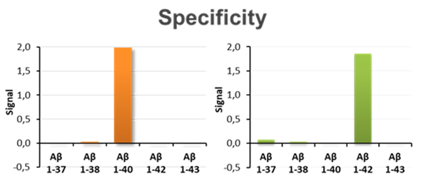
The chart shows the specificity of Aβ40 and Aβ42 determinations characterised by the almost complete absence of cross-reactivity with other peptides similar to each of the target peptides.

CLINICAL RESULTS

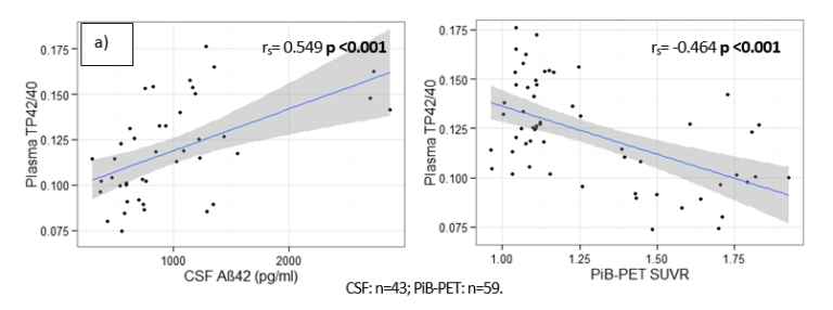
Plasma Aβ42/Aβ40 ratio (TP42/40) values measured by ABtest-IA correlate directly with cerebrospinal fluid Aβ42 levels and inversely with cortical amyloid burden measured by PET scanning. (https://pubmed.ncbi.nlm.nih.gov/31787105)

Plasma Aβ42/Aβ40 ratio predicts brain amyloid burden and can discriminate normal amyloid PET-positive vs. negative individuals with an area under the curve (AUC) of 0.896 among people without symptoms of cognitive impairment. (https://pubmed.ncbi.nlm.nih.gov/32179698).

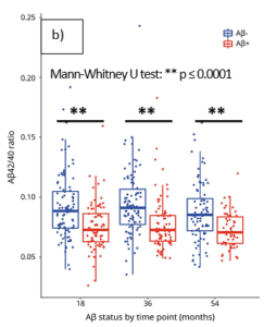
The Aβ42/Aβ40 ratio measured by ABtest-IA is lower in individuals with cortical amyloid burden above the normal level in Amyloid-PET (Aβ+) than in individuals with a burden below the threshold (Aβ-) ( https://pubmed.ncbi.nlm.nih.gov/32179698).

Hypothetical recruitment scenario for a clinical trial

Adaptado de Insel et al. : Assessing risk for preclinical β-amyloid pathology with APOE, cognitive, and demographic information in Alzheimers Dement (Amst). 2016; 4: 76–84. https://www.ncbi.nlm.nih.gov/pmc/articles/PMC5045949.

If recruitment is carried out directly based on PET-amyloid level, 500 scans would be needed whereas if pre-screening of participants on the basis of their plasma Aβ42/Aβ40 levels is implemented, the number of PET scans would be reduced to 208 to confirm the plasma test result.

This two-step screening strategy would reduce the screening failure rate from 70% to ~28%, making participation in clinical trials less demanding for individuals and substantially reducing the length of the screening period and associated costs.

In addition, ABtest-IA allows differentiated determination of the levels of free Aβ40 and Aβ42 fraction in plasma (FP40 and FP42) as well as their total plasma levels (TP40 and TP42).

The free fraction (FP40 and FP42) obtained by our immunoassay is determined from an undiluted sample and represents the fraction of these peptides that is directly available in the sample, free of interactions with other plasma components that prevent their immuno-detection.

The total levels (TP40 and TP42) are obtained from an aliquot of the same sample, diluted in a proprietary buffer so that, together with the free one, the fraction of these peptides that was initially sequestered for the assay due to their interactions with other plasma components is also measured. Overall, the free fraction represents only about 40% of the total peptide quantified (TP42 and TP40).

Our results indicate that in most cases determining the TP42/40 ratio is sufficient for practical purposes regarding disease detection or treatment monitoring. However, in certain research projects the additional assessment of the free fraction of both peptides (FP40, FP42) could provide information about the effect that the plasma matrix of each individual might be exerting on the metabolism and blood Aβ-peptide levels.